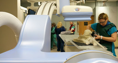
Veterinary Imaging, Opens Up New Doors of Treatment and Prevention for Animals
Veterinary imaging is used to obtain medical images of animal bodies for the diagnosis of chronic diseases. It is a non-invasive method developed from human medicine with the help of diagnostic imaging devices. Veterinary imaging devices help detect sinus or nasal diseases, differentiating neoplasia from rhinitis and therefore guide biopsy examination results. These devices are also used to detect brain hemorrhage and skull fractures. Moreover, the imaging devices allow the visualization of different types of brain tumors. Furthermore, the use of veterinary imaging eliminates the use of photo processing supplies, film jackets, darkroom, and film which reduces the cost of diagnosis.
However, compared to magnetic resonance imaging (MRI), veterinaryimaging is not as sensitive in the case of detecting vascular condition, inflammation
and tumors. Moreover, imaging devices can only be operated by skilled users and
also require a computer support for software and hardware related problems.
Veterinary imaging is a growing area of excellence, opens up new doors of
treatment and prevention for pets. It allows us to detect, plan, and execute
preventative measures for many health problems. Early detection can greatly
impact disease progression and often mean the difference between life and
death.
Veterinary imaging is a growing field of excellence in the
animal health profession. The goal is to contribute to your knowledge of normal
physiology and illness, with the ultimate goal of prevention, diagnosis, and
treatment. The scope of veterinary imaging encompasses research into the
diagnosis and treatment of experimental and clinical disease, as well as the
collection, preparation and analysis of data from specialized images. In the
field of medicine, veterinary radiologists are integral members of the medical
imaging team and play an important role in ensuring that high quality images
are produced.
The veterinary imaging process begins with a mammogram,
which is a form of x-ray that uses gamma rays to create an image of the
abdominal viscera. This allows radiologists to look into the heart, blood
vessels and bones of the animals for assessment. Abnormalities and diseases can
be revealing in these organs and systems, allowing for the proper diagnosis and
treatment. Mammograms are performed in both acute and chronic conditions. As
part of the treatment program, radiologists often use computed tomography (CT)
scanners or magnetic resonance imaging (MRI) to reveal a more detailed picture
of the affected organs and systems.
As advances in technology and modern approaches to
veterinary medicine are introduced, the scope of veterinary imaging will
continue to expand. New technologies and more efficient equipment mean that
radiation therapy, image guided surgery, and other specialized services are
becoming more commonplace. New diagnostic tools, such as mammography, magnetic
resonance imaging, and ultrasound have been developed specifically for oncology
practice. The future of veterinary imaging will continue to challenge the
boundaries of traditional practices in veterinary medicine. The future of
veterinary imaging does not depend on how well it can diagnose and treat
diseases and disorders, but it lies in being able to prevent them. Veterinary
imaging will continue to contribute towards improved health care by detecting
and treating problems before they cause irreversible damage.


Comments
Post a Comment